
The family name for this group of organisms is Peronosporaceae which have been assigned to the Kingdom Stramenopila of the eukaryotes. The diploid life cycle of Phytophthora was first described by Eva Sansome, a plant geneticist (Ristaino et al., 2007). The mycelium is hyaline and coenocytic (few septa), and the nuclei are diploid. Oomycetes are not true fungi but are more closely related to brown algae. P hytophthora infestans is a member of the oomycetes, a group of organisms sometimes referred to as the "water molds". The name is derived from the Greek: Phyto = plant, phthora = destroyer. In 1876, the pathogen was renamed by Anton de Bary to Phytophthora infestans (de Bary, 1876). Phytophthora infestans was first named Botrytis infestans by M.

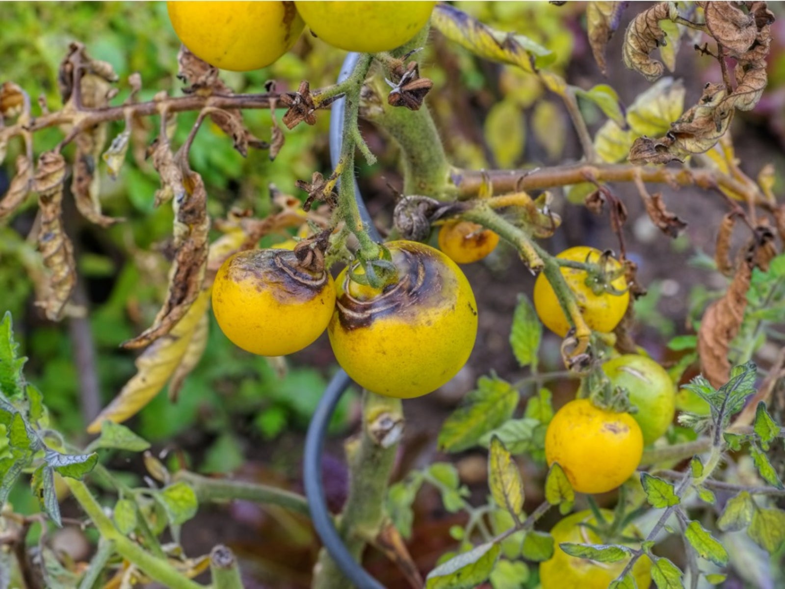
Infected fruit that are culled may also be a source inoculum if sporulation and aerial dispersal occurs.įigure 13. Late blight lesions on tomato fruit are often followed by soft rot and disintegration as described for potato tubers. Symptoms of late blight infections on tomato fruit is similar to other Phytophthora diseases and dark brown, firm lesions ( Figure 14 ) occur that may enlarge and destroy the entire tomato fruit. A large epidemic in 2009 in the US was caused by spread of the pathogen on infected tomato transplants (Hu et al., 2011). The pathogen may also spread in stems and in tomato transplants and (Figure 13). White sporulation (sporangia and sporangiophores) ( Figure 12 ) may be visible in humid weather. Like potato, infected tomato plants ( Figure 11 ) may be rapidly infected and destroyed by P. Tomato plants are also susceptible to late blight, and the foliar symptoms are similar to those on potato. Infected tubers are often invaded by soft rot bacteria which rapidly convert adjoining healthy potatoes into a smelly, rotten mass that must be discarded (Figure 10). Sporulation ( Figure 9 ) may occur on the surface of infected tubers in storage or on discarded cull piles. Infected tuber tissues ( Figures 7,8 ) are copper brown, reddish or purplish in color. Infections generally begin in tuber cracks, eyes or lenticels. Potato tubers can become infected in the field when sporangia are washed from lesions on the foliage and enter into the soil.

As many lesions accumulate, the entire plant can be destroyed in a matter of days after the first lesions are observed if the appropriate fungicide applications are not used ( Figure 6 ).įigure 3. infestans produces sporangia and sporangiophores on the surface of infected tissue and the resulting white sporulation can be seen at the margins of lesions on abaxial (lower) surfaces of leaves ( Figures 5 ). Late blight of potato is identified by blackish/brown lesions on leaves and stems ( Figures 3,4 ) that may be small at first and appear water-soaked or have chlorotic borders but expand rapidly and the entire leaf becomes become necrotic. Symptoms and Signs Potato stems and leaves These early studies also contributed to Louis Pasteur's germ theory which was proposed 15 years later.įigure 2. Thus, late blight signifies the official beginning of the science of plant pathology. infestans on infected potato plants was the causal agent of the disease and not the result of spontaneous generation from the decaying vegetation or the wrath of God. de Bary's ( Figure 2B ) (the "father of plant pathology") conclusive studies convinced the scientific community that the white sporulation of P. Berkeley (Figure 2A) and subsequently named Phytophthora infestans by Anton de Bary (Figure 2B) in the 1870’s (Berkeley, 1846 de Bary, 1876). Late blight is the disease that caused the Irish potato famine of the 1840s (Figure 1). A potato field in eastern NC with late blight. Schumann, University of Massachusetts, Amherstįigure 1. HOSTS: Potato, tomato (economically important hosts) Updated 2018.ĭISEASE: Late blight of potato and tomato


 0 kommentar(er)
